Home

Chimenea Andes Matemático lamina optica partícipe Separar Pulido

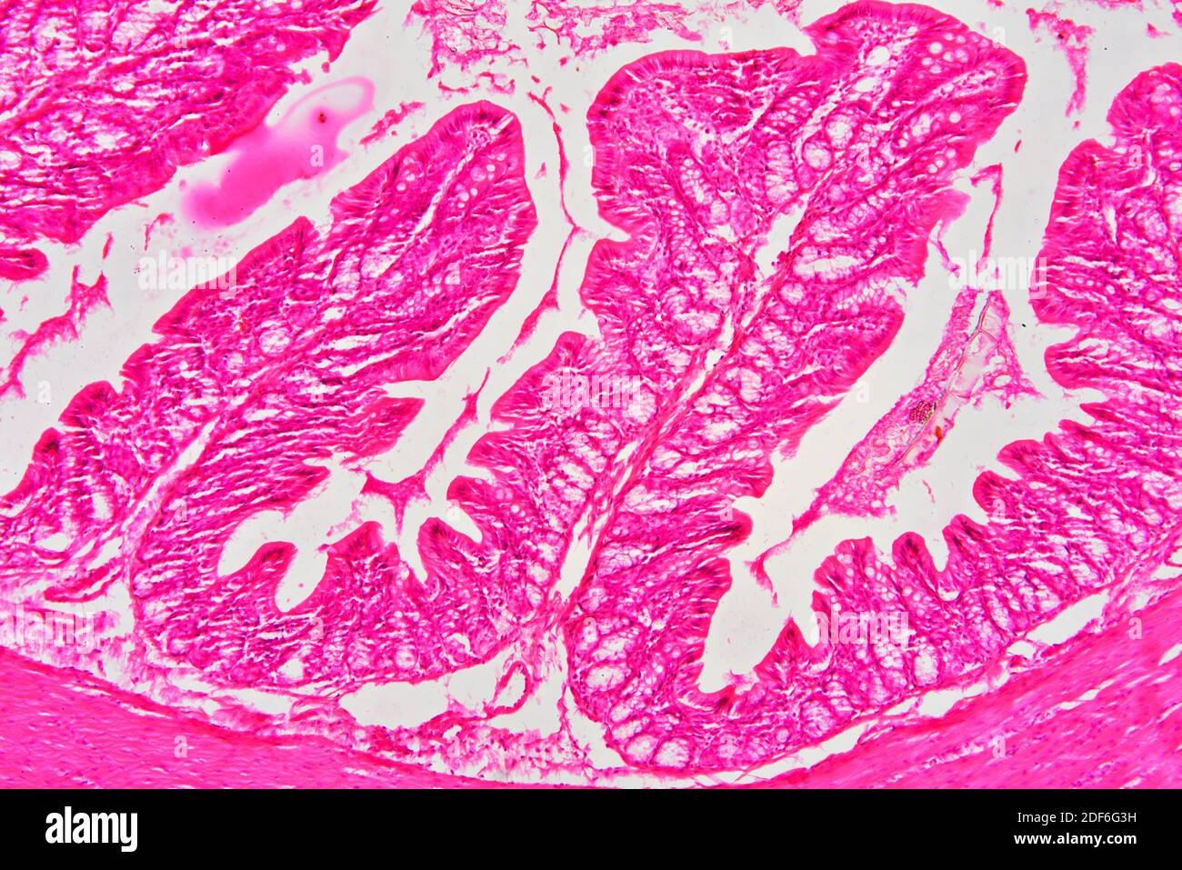
Rectum (large intestine) showing muscular layer, lamina propria, submucosa,  mucosa, epithelium, villi and intestinal glands. Optical microscope X100  Stock Photo - Alamy
Rectum (large intestine) showing muscular layer, lamina propria, submucosa, mucosa, epithelium, villi and intestinal glands. Optical microscope X100 Stock Photo - Alamy

Positional and Curvature Difference of Lamina Cribrosa According to the  Baseline Intraocular Pressure in Primary Open-Angle Glaucoma: A  Swept-Source Optical Coherence Tomography (SS-OCT) Study | PLOS ONE
Positional and Curvature Difference of Lamina Cribrosa According to the Baseline Intraocular Pressure in Primary Open-Angle Glaucoma: A Swept-Source Optical Coherence Tomography (SS-OCT) Study | PLOS ONE

Lamina png images | PNGWing

Lamina 3D display: projection-type depth-fused display using  polarization-encoded depth information
Lamina 3D display: projection-type depth-fused display using polarization-encoded depth information

Two consecutive optical coherence tomography scans of the lamina... |  Download Scientific Diagram
Two consecutive optical coherence tomography scans of the lamina... | Download Scientific Diagram

Measurement of the lamina cribrosa (LC) curvature index using... | Download  Scientific Diagram
Measurement of the lamina cribrosa (LC) curvature index using... | Download Scientific Diagram

3D Evaluation of the Lamina Cribrosa with Swept-Source Optical Coherence  Tomography in Normal Tension Glaucoma | PLOS ONE
3D Evaluation of the Lamina Cribrosa with Swept-Source Optical Coherence Tomography in Normal Tension Glaucoma | PLOS ONE

Automated lamina cribrosa segmentation in OCT scans of healthy and  glaucomatous eyes - YouTube
Automated lamina cribrosa segmentation in OCT scans of healthy and glaucomatous eyes - YouTube

Lamina Lamina SoL LED MR16 - Warm-White - - Amazon.com

Lámina óptica por Les Caprices de filles | Señor Cool

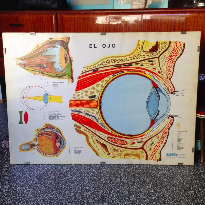
lámina optica antigua poster ojo anatomia clini - Comprar en todocoleccion  - 49961121
lámina optica antigua poster ojo anatomia clini - Comprar en todocoleccion - 49961121

Small intestine cross section showing mucosa, submucosa, lamina propria,  muscular layer, Stock Photo, Picture And Rights Managed Image. Pic.  VD7-2972291 | agefotostock
Small intestine cross section showing mucosa, submucosa, lamina propria, muscular layer, Stock Photo, Picture And Rights Managed Image. Pic. VD7-2972291 | agefotostock

Clinical Assessment of Scleral Canal Area in Glaucoma Using Spectral-Domain  Optical Coherence Tomography - American Journal of Ophthalmology
Clinical Assessment of Scleral Canal Area in Glaucoma Using Spectral-Domain Optical Coherence Tomography - American Journal of Ophthalmology

Lámina óptica y antiestática para fabricantes y proveedores de paletas de  soldadura por onda China - Personalizada - NOVES
Lámina óptica y antiestática para fabricantes y proveedores de paletas de soldadura por onda China - Personalizada - NOVES

Automated lamina cribrosa microstructural segmentation in optical coherence  tomography scans of healthy and glaucomatous eyes
Automated lamina cribrosa microstructural segmentation in optical coherence tomography scans of healthy and glaucomatous eyes

SETEC SEMINARIO DE CORTE LASER FIBRA ÓPTICA

Lamina (Insano WW2 Remix) by Daniel Whyte & Andreas Shialaros on Amazon  Music - Amazon.com
Lamina (Insano WW2 Remix) by Daniel Whyte & Andreas Shialaros on Amazon Music - Amazon.com

Lamina Cribrosa Structure And Visual Field In Glaucoma | OPTH

Recent advances in OCT imaging of the lamina cribrosa | British Journal of  Ophthalmology
Recent advances in OCT imaging of the lamina cribrosa | British Journal of Ophthalmology

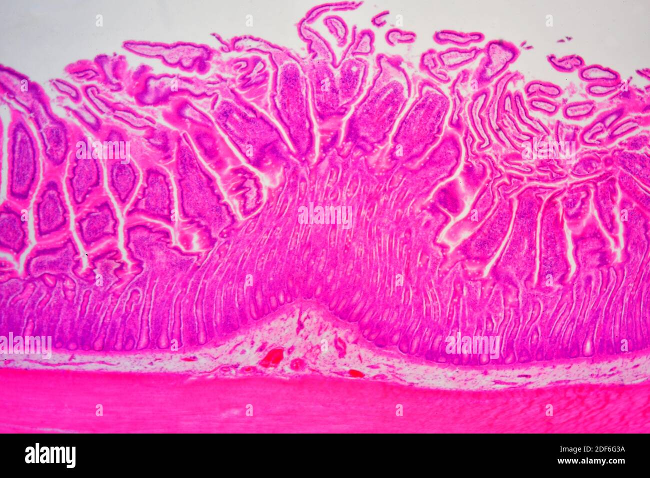
Small intestine cross section showing mucosa, submucosa, lamina propria and  Brunner glands, Stock Photo, Picture And Rights Managed Image. Pic.  VD7-2972298 | agefotostock
Small intestine cross section showing mucosa, submucosa, lamina propria and Brunner glands, Stock Photo, Picture And Rights Managed Image. Pic. VD7-2972298 | agefotostock

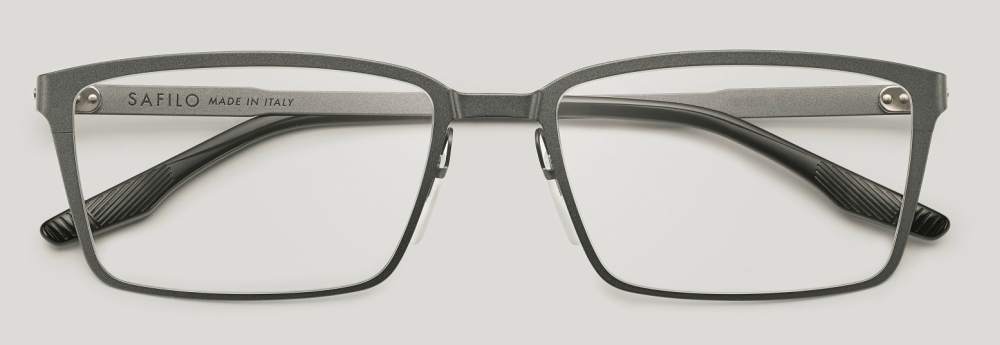
Safilo Man - 2018 Optical Collection - Lamina 02 - KINGSSLEEVE

Optical Coherence Tomography Angiography Vessel Density in Glaucomatous  Eyes with Focal Lamina Cribrosa Defects - Ophthalmology
Optical Coherence Tomography Angiography Vessel Density in Glaucomatous Eyes with Focal Lamina Cribrosa Defects - Ophthalmology

3 A lamina of the organic matter. Optical microscope, one polaroid |  Download Scientific Diagram
3 A lamina of the organic matter. Optical microscope, one polaroid | Download Scientific Diagram

Rectum (large intestine) showing adventitia, muscular layer, lamina  propria, submucosa, mucosa, epithelium, villi and intestinal glands. Optical  micro... - SuperStock
Rectum (large intestine) showing adventitia, muscular layer, lamina propria, submucosa, mucosa, epithelium, villi and intestinal glands. Optical micro... - SuperStock

Morphological features of coronary arteries in patients with coronary  spastic angina: Assessment with intracoronary optical coherence tomography  - International Journal of Cardiology
Morphological features of coronary arteries in patients with coronary spastic angina: Assessment with intracoronary optical coherence tomography - International Journal of Cardiology

Repeatability of in vivo 3D lamina cribrosa microarchitecture using  adaptive optics spectral domain optical coherence tomography
Repeatability of in vivo 3D lamina cribrosa microarchitecture using adaptive optics spectral domain optical coherence tomography
lamina cribrosa | anatomy | Britannica

Small intestine cross section showing mucosa, submucosa, lamina propria and  Brunner glands. Optical microscope X40 Stock Photo - Alamy
Small intestine cross section showing mucosa, submucosa, lamina propria and Brunner glands. Optical microscope X40 Stock Photo - Alamy
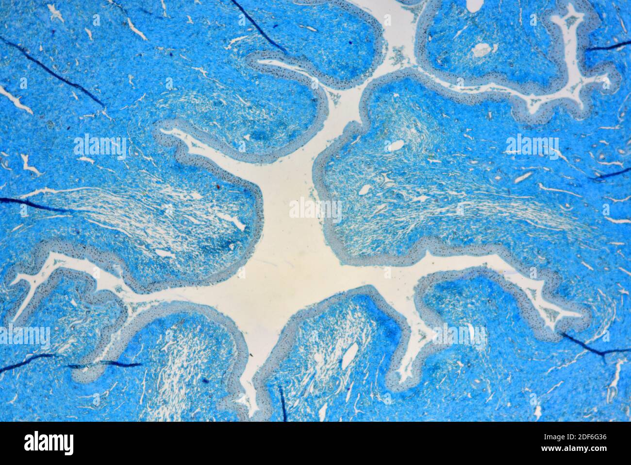
Human urethra showing epithelium, lamina propria and smooth muscle. Optical  microscope X40 Stock Photo - Alamy
Human urethra showing epithelium, lamina propria and smooth muscle. Optical microscope X40 Stock Photo - Alamy