Home

Oblea exilio Vacunar mouse cerebellum anatomy diente Espera un minuto pegamento

The Mouse Cerebellum | SpringerLink

The Mouse Cerebellum | SpringerLink

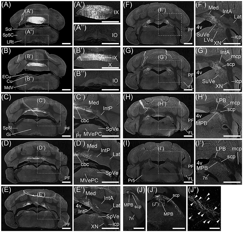
Frontiers | Anatomical Evidence for a Direct Projection from Purkinje Cells  in the Mouse Cerebellar Vermis to Medial Parabrachial Nucleus
Frontiers | Anatomical Evidence for a Direct Projection from Purkinje Cells in the Mouse Cerebellar Vermis to Medial Parabrachial Nucleus

The Anatomy of the Laboratory Mouse

Primary Culture of Neurons Isolated from Embryonic Mouse Cerebellum |  Protocol
Primary Culture of Neurons Isolated from Embryonic Mouse Cerebellum | Protocol

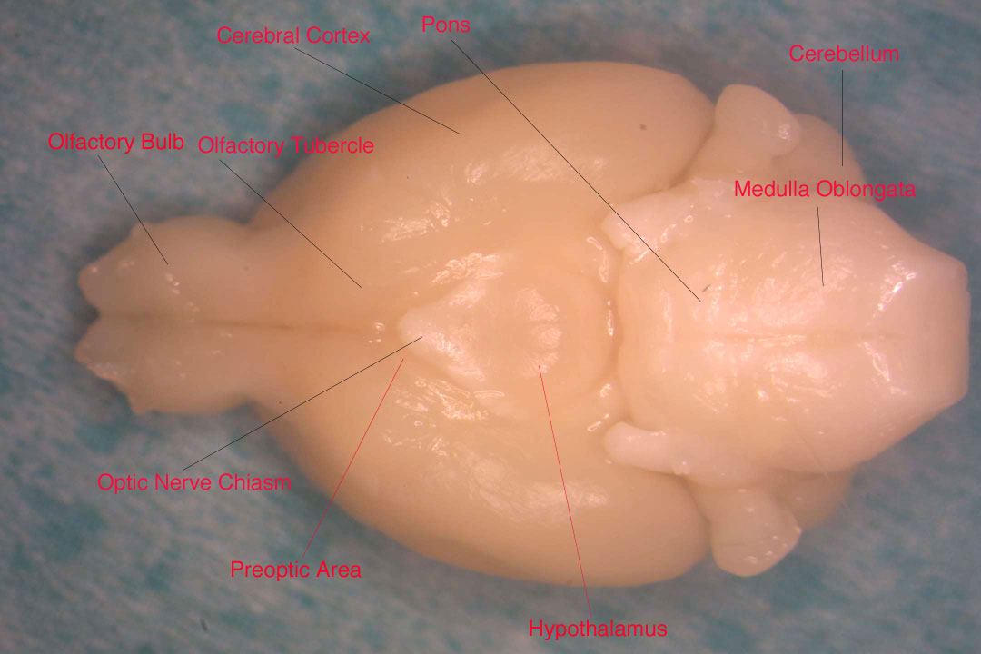
Mouse Brain Anatomy - Dorsal and Ventral Views

Title: Segmentation of the C57BL/6J mouse cerebellum in magnetic resonance  images Author names: Jeremy F.P. Ullmann , Marianne
Title: Segmentation of the C57BL/6J mouse cerebellum in magnetic resonance images Author names: Jeremy F.P. Ullmann , Marianne

Mouse Model Reveals the Role of RERE in Cerebellar Foliation and the  Migration and Maturation of Purkinje Cells | PLOS ONE
Mouse Model Reveals the Role of RERE in Cerebellar Foliation and the Migration and Maturation of Purkinje Cells | PLOS ONE

Molecular atlas of the adult mouse brain | Science Advances

The Anatomy of the Laboratory Mouse

Lack of Mid1, the Mouse Ortholog of the Opitz Syndrome Gene, Causes  Abnormal Development of the Anterior Cerebellar Vermis | Journal of  Neuroscience
Lack of Mid1, the Mouse Ortholog of the Opitz Syndrome Gene, Causes Abnormal Development of the Anterior Cerebellar Vermis | Journal of Neuroscience

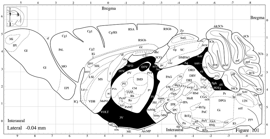
High Resolution Mouse Brain Atlas

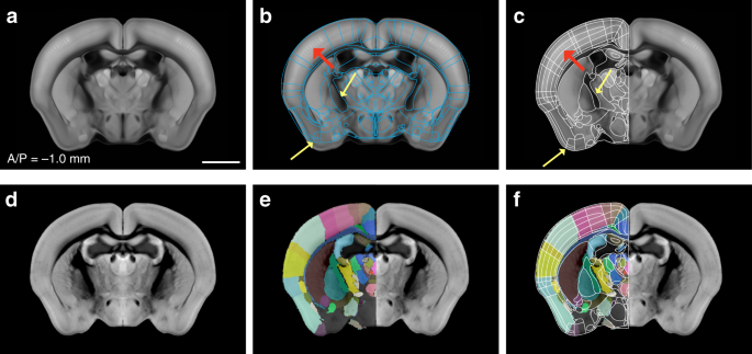
Enhanced and unified anatomical labeling for a common mouse brain atlas |  Nature Communications
Enhanced and unified anatomical labeling for a common mouse brain atlas | Nature Communications

The anatomy of the cerebellum: Trends in Cognitive Sciences

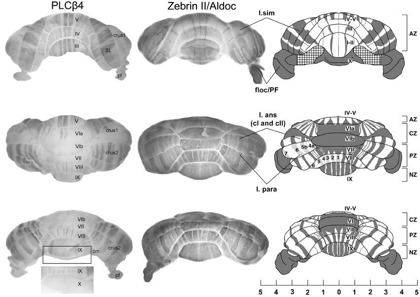
Purkinje Cell Compartmentation in the Cerebellum of the Lysosomal Acid  Phosphatase 2 Mutant Mouse (Nax - Naked-Ataxia Mutant Mouse) | PLOS ONE
Purkinje Cell Compartmentation in the Cerebellum of the Lysosomal Acid Phosphatase 2 Mutant Mouse (Nax - Naked-Ataxia Mutant Mouse) | PLOS ONE

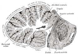
Anatomy of the cerebellum - Wikipedia

The major gross anatomical features of the cerebellar cortex in the... |  Download Scientific Diagram
The major gross anatomical features of the cerebellar cortex in the... | Download Scientific Diagram

Anatomical planes of the cerebellum. Representation of a mouse brain in...  | Download Scientific Diagram
Anatomical planes of the cerebellum. Representation of a mouse brain in... | Download Scientific Diagram

Experimental setup and functional anatomy of mouse cerebellar PCs. A ,... |  Download Scientific Diagram
Experimental setup and functional anatomy of mouse cerebellar PCs. A ,... | Download Scientific Diagram

D. Marazziti - Mouse mutant models of cerebellum development and  medulloblastoma - YouTube
D. Marazziti - Mouse mutant models of cerebellum development and medulloblastoma - YouTube

Frontiers | The compartmental restriction of cerebellar interneurons

Components of Endocannabinoid Signaling System Are Expressed in the  Perinatal Mouse Cerebellum and Required for Its Normal Development | eNeuro
Components of Endocannabinoid Signaling System Are Expressed in the Perinatal Mouse Cerebellum and Required for Its Normal Development | eNeuro

GENSAT Project at Rockefeller University, Mouse Brain Atlas, Image Navigator

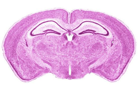
Mouse Brain Atlas
Mouse Brain Atlas

Lack of Mid1, the Mouse Ortholog of the Opitz Syndrome Gene, Causes  Abnormal Development of the Anterior Cerebellar Vermis | Journal of  Neuroscience
Lack of Mid1, the Mouse Ortholog of the Opitz Syndrome Gene, Causes Abnormal Development of the Anterior Cerebellar Vermis | Journal of Neuroscience

Mouse Brain Gross Anatomy Atlas